ADVANCED
NANO-MATERIALS
STRUCTURES AND
APPLICATIONS
ANMSA focuses on design, engineering and manipulation of novel nanocomposite and hybrid materials and structures on the micron and nanometric scale for a broad range of applications in for instance, microelectronics, diagnostics and photovoltaic cells. The control of patterns on sub-micrometre lateral length scales is of considerable technological interest and is especially relevant for tailoring the properties of novel functional materials. The growing demand for better performance, reduced energy consumption, high-throughput and higher levels of complexity and integration raise the need for alternative technologies capable of more than the generation or reproduction of patterns below the sub-100 nm to develop tunable 3D structures and thus, put these topics among the priorities of our research agenda. We are developing and implementing advanced, cost-effective soft lithography techniques and particularly, electrohydrodynamic (EHD) patterning. We also explore and exploit bottom-up and top-down fabrication strategies to create new classes of high performance structures to mimic the intriguing hierarchical morphologies and patterns found in nature.
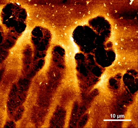
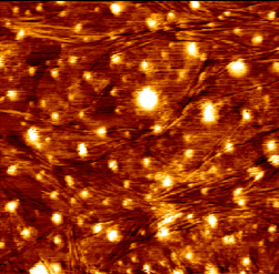
A paradigm shift from bulky and expensive diagnostics to rapid real-time chemical and biological detection is offered by micro-engineering technology with enhanced functions and performance combining sample preparation, manipulation and delivery. Medical diagnostics, environmental monitoring, homeland security and forensics increasingly demand specific and field-deployable analytical technologies for quick point-of-care diagnostics. Enhancement via localized optical fields on nanoscale metallic materials generates huge signals in SERS, enabling single molecule detection. This enhancement can be tuned by manipulation of the surface roughness and architecture at the sub-micron level. Simultaneous identification of different molecules in a mixture is among the distinct advantages of SERS over other spectroscopic techniques. This project is focussed on development of reproducible, well-defined and stable surfaces for SERS detection in a microfluidic device. These are reliably produced, tested and integrated into a fluidic chip with optical fibre based SERS detection. Here we combine high sensitivity offered by SERS surfaces produced by optimised electrohydrodynamic lithography to deliver a completely portable miniaturised SERS-based system for point-of-care diagnostics.
[1] Nie, S., Emory, S. R. Science 1997, 275, 1102.[2] Ozbay, E. Science 2006, 311, 189.
[3] Lin, M. et al., J. Food Sci. 2008, 73, 129.
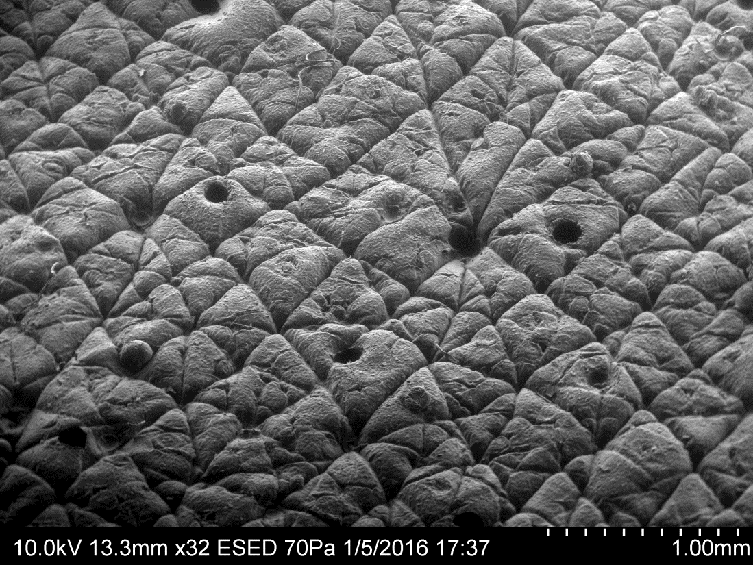
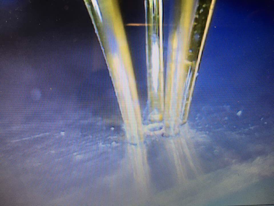
Most skin care products nowadays contain active moisturizing agents that moisturize the skin leaving it with a healthy and smooth appearance. This project is co-funded with P&G and aims to study the retention processes on the human skin after the application of such products to better understand the underlying mechanisms and the way we can change parameters in the formulation of skin care products in order to improve their effectiveness. The representative formulations include body wash products and the moisturizing agent is petrolatum, which has been proved to be the most effective moisturizing element, including its occlusive properties that diminish trans-epidermal water loss and its protective capabilities to the skin from wind and dryness. In this project we are recreating shower conditions in a controlled, laboratory environment, where we can test the products on novel optimised skin mimics and then study those for the retention of actives. This study also required the production of dedicated representative emulsions and a water jet set-up specifically esigned and used to remove the emulsion from the skin mimics.
[1]. D. S. Morrison, Petrolatum: A Useful Classic, Cosmetics & Toiletries magazine Vol. 111, 1996, 59.[2]. A.O. Barel, M. Paye, H.I. Maibach, Handbook of Cosmetic Science and Technology, 3rd Edition.
A DSTL funded project focusing on synthesizing novel 3D-nanomaterial structures utilizing a synthetic biology approach to develop surfaces capable of optical and photonic characteristics. Synthetic biology mimics processes seen in nature and exploiting them to yield real world applications of ‘smart materials’. One of the main areas of focus is on the development of a paper-based bio-detector for rapid colorimetric detection of heavy metals, such as Hg2+ and Pb2+ that can be visualized with the naked-eye.
Human progress has been punctuated by the development of new materials that have revolutionised the way we live. The transition from bronze to iron ages catalysed a sea change in agriculture while the advent of silicon semiconductors brought on the new information age. In this project we aim to explore a whole range of new materials produceControlling Collagen Formation in Wounds Using Smart Dressingsd by combining the exquisite morphological conformaty offered by biological materials (in this case bacteriophage) with advances in material sciences. The aim of this combination of disciplines (a core part of Synthetic Biology) is to generate new optoelectronic materials that will provide opportunities across the defence sector. Nanostructures with periodicities that are comparable to the light wavelength, i.e., photonic band-gap (PBG) and meta-materials, can trap certain colours enabling control of light to achieve amazing optical effects that are impossible with conventional optics. One of the main obstacles to realize these concepts is the enormous difficulty to fabricate periodic three-dimensional (3D) nanostructures. This project makes use of viruses to construct such 3D nano-architectures. Viral fragments are used similar LEGO bricks, which correctly fit together in one unique way. The viral blocks are designed to build-up well-defined assembled 3D-periodic network nanoscale structures which exhibit unique optical properties. These new developments of tailorable platforms realizes the concept of changing the shape and spacing of internal structures within novel biosynthetically generated metamaterials, opening the door to transforming light in many ways producing tailorable, low-observable devices and materials that could be applied to large surface areas.
[1]. Guiding light with conformal transformations. N. I. Landy and W. J. Padilla, Opt. Express, 2009, 17(17), 14872–14879.[2]. Photonic crystals in the optical regime: past, present and future. T.F. Krauss, R.M. De La Rue, Prog. Quantum Electron. 1999, 23, 51 96.
[3]. Self-assembly approach to optical metamaterials. J.F. Galisteo, et al. J. Opt. A: Pure Appl. Opt. 2005, 7, S244–S254.

Traumatic Brain Injury (TBI) is a leading cause of morbidity and mortality worldwide with neuro-disabilities requiring long-term care. Timely TBI-diagnosis is at the heart of national guidelines, however, this is poorly supported by urrent technologies which fall short of the PoC diagnostic needs [1]. This project aims to enable an approach based on Raman spectroscopy of the back of the eye, capable of detecting changes in CSF without the need for a lumbar puncture or expensive radiological scans. It is envisioned that Raman spectroscopy of the optic nerve could be used to observe subtle chemical changes as a result of TBI that are otherwise undetectable in the peripheral circulation [2, 3].
[1] National Audit Office. London: The Stationery Office., 2010.[2] Igor V. Ermakov et al. Optics Letters, 2001.
[3] Marro M. et al Journal of Biophotonics, 2014.
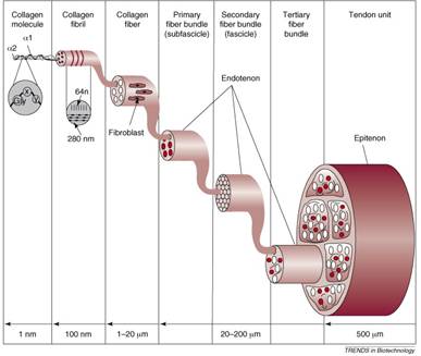
Collagen networks determine the functional properties of most connective tissue. Through self-assembly of procollagen, followed by covalent cross-links, a network of fibres is established in a hierarchical fashion, across molecular and macro-scales. Characteristic features of this network influences cell growth and therefore tissue function. [1] For example, the 15nm spacing of the collagen lattice within the cornea causes scattered light to interfere destructively resulting in its transparency. [2] Scarring within tissue is the result of a disrupted collagen matrix, with the tissue only ever returning to 70% strength, inducing discomfort and even blindness for a patient. [3] Collagen self-assembly is both a physical and chemical process. Therefore, identification of methods to manipulate the formation of collagen fibrils in a controlled manner will contribute towards the development of a new generation of dressings for traumatic soft tissue injury patients. Manipulation can be undertaken through changes in topographical surface structure, the introduction of inorganic ions and the presence of an electric field. Through matrix manipulation the development of life altering scarring can be reduced.
[1] Collagen Structure and mechanics Peter Fratzl, Springer, (2008), USA, pages 10-20.[2] Connon, C.J., Meek, K.M., Organization of corneal collagen fibrils during the healing of trephined wounds in rabbits, Wound Repair Regeneration, (2003), 11:(71-78).
[3] Richard Clark, The Molecular and Cellular Biology of Wound Repair, Springer Science, Second Edition, 2012.
[4] Liu, Y., Ramanath, H.S., Wang, D-A., Tendon tissue engineering using scaffold enhancing strategies, Trends in Biotechnology, (2008), 26:4(201-209).